Seeing Inside the Bone with a 3D Cone CT Scan
What Is Cone-Beam Computed Tomography?
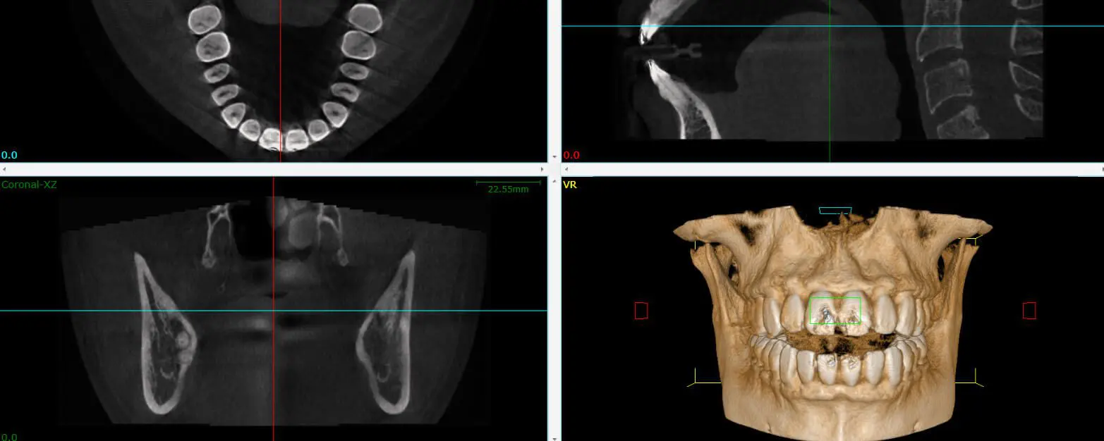
Cone-beam CT creates a 3D view and cross-sections of the target area. The information it provides is critical for a number of situations, including placing dental implants, performing a complex root canal or extraction, or searching for the source of pain or infection. CBCT gives us the information we need about your condition before a dental procedure.
The Difference Between CBCT and a Hospital CT or CAT Scan
Dental cone-beam CT emits less radiation and provides a more complete picture than CT and CAT scans. Hospital CTs work by taking a series of parallel X-ray images of the head, from top to bottom. But what about the areas in between those images? One of the biggest drawbacks of these kinds of scans is that they don’t see everything. They have to fill in the gaps with the computer’s educated guesses. Another drawback is that taking so many X-rays exposes the patient to more radiation.
Unlike with CT and CAT scans, the cone-beam CT scanner circles the head, ensuring that the images it takes overlap each other, leaving no gaps. The radiation is also much weaker. The most radiation hits the area of interest, which is where the images overlap to construct the 3D model. This is how CBCT is able to provide a more complete image with less radiation exposure.
How Do I Benefit from Cone-Beam 3D Imaging?
Aside from providing more complete information, dental CBCT scans emit very little radiation, equivalent to just one day of normal background radiation. The risk of such a low level of radiation is much less than that of getting an inaccurate diagnosis. A CBCT scan ensures that Dr. Paymon will be able to diagnose you with the best possible accuracy. For more information about cone-beam 3D imaging, give us a call at 360-352-6399 or send us an email.